We built identifyamyloid.com — an illustrated study of the amyloid heart — because the single biggest obstacle to treating cardiac amyloidosis is recognizing it in the first place. The disease is treatable and more common than most clinicians expect, yet it is routinely mistaken for ordinary heart failure. This post walks through the key points; the full drawings, 3-D models, and sources live on the site.
What it is
Cardiac amyloidosis is an infiltrative cardiomyopathy. Misfolded protein deposits in the space between heart-muscle cells; the walls thicken, the ventricle stiffens, filling pressures rise, and the result is a restrictive cardiomyopathy that drives atrial fibrillation, fluid retention, and heart failure. More than 98% of cardiac amyloidosis comes from just two precursor proteins: transthyretin (ATTR), where a liver-made transport protein destabilizes and misfolds, and immunoglobulin light chains (AL), where a plasma-cell clone releases misfolding light chains.
The distinction matters because the treatments are completely different — ATTR is addressed with transthyretin stabilizers and gene silencers, AL with therapy directed at the plasma-cell clone — and because both are now treatable in ways they were not a decade ago. That is exactly why finding these patients early has become urgent.
Why it hides
Because the process is restrictive, the heart stiffens before it weakens: the ejection fraction usually looks preserved until late in the disease. A study can read as "normal" or as heart failure with preserved ejection fraction, and the patient is never worked up for amyloid. The signatures are there on the echo — increased wall thickness out of proportion to any history of hypertension, a restrictive mitral-inflow pattern, tissue-Doppler velocities below 5 cm/s, and the classic relative apical sparing on a longitudinal-strain bullseye — but they are easy to pass over in a busy lab.
Clues that should raise suspicion
Increased LV wall thickness without a hypertensive history · low ECG voltage discordant with wall thickness · bilateral carpal tunnel syndrome (often 5–9 years earlier) · lumbar spinal stenosis · spontaneous biceps-tendon rupture · intolerance to antihypertensive or heart-failure therapy · low-flow, low-gradient aortic stenosis · a restrictive filling pattern with apical sparing on strain.
How to diagnose it
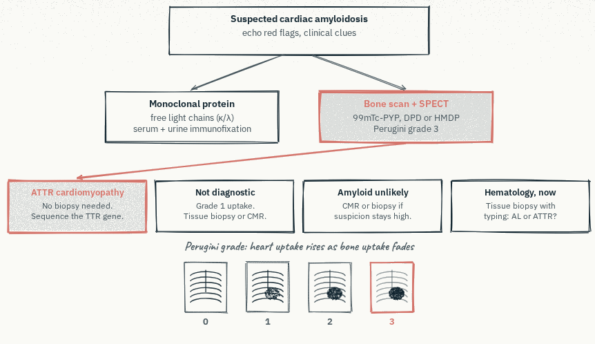
The modern work-up is largely non-invasive and rests on two pillars done together:
- Screen for a monoclonal protein — serum free light chains plus serum and urine immunofixation — to look for the light chains of AL.
- Bone scintigraphy — a Tc-99m PYP/DPD/HMDP scan with SPECT — which takes up tracer avidly in ATTR cardiac amyloidosis.
Read together, these two tests resolve into a small set of combinations: strong cardiac tracer uptake with no monoclonal protein can confirm ATTR cardiac amyloidosis without a biopsy, while any positive monoclonal-protein result sends the patient toward hematology and tissue confirmation for AL. An endomyocardial or fat-pad biopsy settles the ambiguous cases.
Why early detection matters
Delay is the disease's defining feature. Patients accumulate years of clues before anyone connects them: carpal tunnel syndrome can precede the heart findings by half a decade, and by the time the heart is implicated, the window in which today's therapies do the most good is already narrowing.
The encouraging corollary is that the opportunity is already in hand. Most of these patients have had an echocardiogram — often the first look at the heart once symptoms appear — and the evidence of amyloid is frequently sitting in that study, waiting to be read.
Where echo AI fits
This is the gap InVision Precision Cardiac Amyloid is built for. It is augmentative AI that reads a routine transthoracic echocardiogram: a segmentation network measures wall thickness on the parasternal long-axis view, and a second network reads myocardial texture and motion across apical-four-chamber frames, giving one result per study. When a study looks suggestive of cardiac amyloidosis, the interpreting physician is alerted to consider referral for confirmatory testing — the signal is surfaced, and the physician decides what happens next. No new scan, no change to how the echo is acquired.
Why a high positive predictive value matters
Cardiac amyloidosis is still uncommon on any given day in the echo lab, and that is exactly what makes specificity — and the positive predictive value (PPV) that follows from it — decisive. When a condition is rare, even a small false-positive rate produces far more false alarms than true ones, and every false flag becomes a bone scan, a hematology referral, or a patient worked up for a disease they do not have.
A model can always be tuned to catch a few more cases by flagging more studies — but at a lower specificity, almost all of those extra flags are false. The comparison below makes the trade-off concrete. Across 1,000 echoes at 1% prevalence, InVision PCA — specificity 99.0% with sensitivity 60.7% in its 510(k) validation4 — flags 16 studies, of which 6 are truly amyloid, with just 10 false flags. Two higher-sensitivity operating points flag 110 and 134 studies to catch one or two more cases, and generate 102 and 125 false flags doing it.
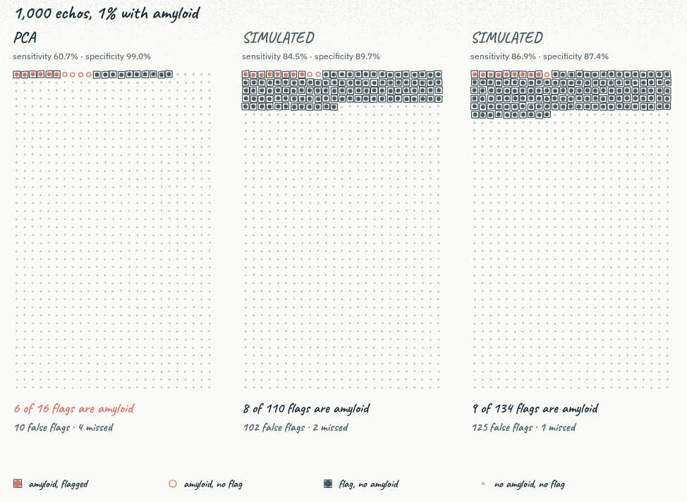
PPV climbs with prevalence, so in a higher-risk population the numbers improve further — in the 510(k) data, PPV rises from 38.4% at 1% prevalence to 55.8% at 2% and 76.5% at 5%.4 And in the one published head-to-head evaluation, in a heart-failure cohort, InVision PCA delivered nearly twice the PPV of another FDA-cleared echo-AI model at each developer's threshold — fewer false positives, at a lower sensitivity.5 For a workflow layered on echoes you already perform, that is the difference between surfacing real cases and burying the lab in false alarms.
See the full study
identifyamyloid.com is a free, illustrated walk-through of the amyloid heart — the protein cascade, the myocardium under the microscope, a 3-D heart as amyloid accumulates, the echo views and red flags, and the full non-invasive work-up — with sources throughout. Share it with your team.